
Immunostaining of PFA fixed 3T3 cells expressing a TOM70-mCherry reporter protein with FluoTag®-X4 Atto 647N anti-RFP sdAb (Cat. No. N0404, dilution 1:500, the mCherry signal is represented in red, the corresponding FluoTag-signal is represented in green and the merge of both channels is represented in yellow). Nuclei were visualized by DAPI staining (blue).

A single motoneuron in the Drosophila larval central nervous system, fixed with 2% PFA. Cell membrane (RFP,red) and synaptic connections (YPet, blue), were stained with FluoTag-X4 anti-RFP (N0404-AF568-L) and FluoTag-X4 anti-GFP (N0304-AT488).
Image credit: Tom Pettini, Matthias Landgraf Lab, University of Cambridge, UK

PFA-fixed Cos7 cells expressing a TOM70-nfRFP-BFP fusion protein (nf: non-fluorescent) were stained with FluoTag®-X4 anti-RFP coupled to Alexa Fluor 647 (Cat. No. N0404-AF647, dilution 1:500). A Greyscale image of the staining performed with N0404-AF647. B False color representation of the image shown in A is displayed in magenta (coloring according to the excitation wavelength of the employed fluorophore). C The corresponding DAPI signal of the depicted section. D Merge of A and C. False color representation of A in magenta and C in blue.

PFA-fixed Cos7 cells expressing a TOM70-nfRFP-BFP fusion protein (nf: non-fluorescent) were stained with FluoTag®-X4 anti-RFP coupled to AZDye 568 (Cat. No. N0404-AF568, dilution 1:500). A Greyscale image of the staining performed with N0404-AF568. B False color representation of the image shown in A is displayed in red (coloring according to the excitation wavelength of the employed fluorophore). C The corresponding DAPI signal of the depicted section. D Merge of A and C. False color representation of A in red and C in blue.
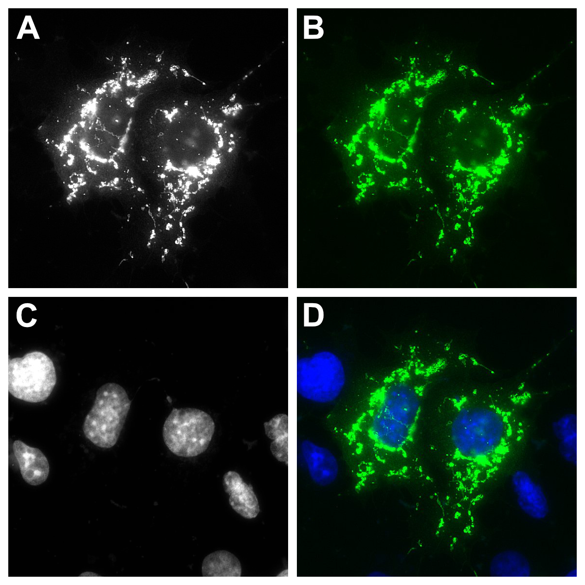
PFA-fixed Cos7 cells expressing a TOM70-nfRFP-BFP fusion protein (nf: non-fluorescent) were stained with FluoTag®-X4 anti-RFP coupled to Atto 488 (Cat. No. N0404-At488, dilution 1:500). A Greyscale image of the staining performed with N0404-At488. B False color representation of the image shown in A is displayed in green (coloring according to the excitation wavelength of the employed fluorophore). C The corresponding DAPI signal of the depicted section. D Merge of A and C. False color representation of A in green and C in blue.


FluoTag®-X4 anti-RFP
400,00 €
FluoTag®-X4 anti-RFP is a blend of two in-house developed single-domain antibodies (sdAbs) recognizing the most common red fluorescent proteins like mRFP, mCherry, DsRed, and other DsRed derivatives with high affinity and specificity.
One of the first red-emitting fluorescent proteins described was DsRed from Discosoma sp. After a couple of years from its publication in 1999 and more than 20 mutations, a monomeric version with several enhanced characteristics appeared as mRFP1. From this promising monomeric red-FP, dozens of monomeric variants have been described, being mCherry one of the most popular derivatives. Today more than 800 entries of various fluorescent proteins can be found in an open-source database with the most currently available variants in the fluorescence protein database “fpbase”
Our nanobodies bind specifically and strongly to DsRed, RFP, and mCherry, among other family members. Look at our specificity chart in the resource section.
FluoTags are directly conjugated to fluorophores, however, they can be equipped with a single fluorophore for more quantitative readouts (FluoTag-Q), with two fluorophores per single-domain antibody (FluoTag-X2), and we also developed a blend of two sdAbs bindings simultaneously the target proteins and each bearing two fluorophores, decorating the target protein with 4 fluorophores in total (FluoTag-X4). For more detailed information on the FluoTags, please check our Technology Section.
| Variations: |
|
||||||||||||||||||||||||
|---|---|---|---|---|---|---|---|---|---|---|---|---|---|---|---|---|---|---|---|---|---|---|---|---|---|
| Related Products: | - | ||||||||||||||||||||||||
| Clone: | 2B12, 2A1 | ||||||||||||||||||||||||
| Host: | Alpaca | ||||||||||||||||||||||||
| Produced in: | E.coli | ||||||||||||||||||||||||
| Application: | IF | ||||||||||||||||||||||||
| Dilution: | 1:250 (corresponding to 5 nM for each sdAb clone) | ||||||||||||||||||||||||
| Capacity: | N/A | ||||||||||||||||||||||||
| Antigen: | - | ||||||||||||||||||||||||
| Targets: | mRFP | ||||||||||||||||||||||||
| Specificity: | mRFP (red fluorescent protein) and other derivatives like mOrange, dsRed1, dsRED2, tdTomato, mCherry and mScarlet-i. | ||||||||||||||||||||||||
| Formulation: |
A mixture of two sdAb clones lyophilized from PBS pH 7.4 containing 2% BSA (US-Origin). Reconstitute with 200 µL of 50 % glycerol in deionized water. We recommend including 0.1 % sodium azide as a preservative if applicable. When reconstituted in 200 µl, the concentration of each individual single-domain antibody is 1.25 µM |
||||||||||||||||||||||||
| kDa: | - | ||||||||||||||||||||||||
| Ext Coef: | - | ||||||||||||||||||||||||
| Shipping: | Ambient temperature | ||||||||||||||||||||||||
| Storing: |
Vials containing lyophilized protein can be stored at 4 °C for 6 months. We recommend reconstituting the protein with 50 % sterile glycerol including 0.1 % sodium azide as preservative if applicable. Minimize the number of freeze-thaw cycles by aliquoting the reconstituted protein. Long term storage at -80 °C for up to 6 months. Working aliquots can be stored at -20 °C for up to 4 weeks. We do not recommend storing the reconstituted protein at 4 °C. |
||||||||||||||||||||||||
| Protocols: |
This Product is not recommended to be used to detect proteins in Western Blott, sdAbs tend to recognize mainly native/folded proteins. Look at detailed protocols and our specificity chart in our Resource Section.
|
||||||||||||||||||||||||
| References: |
|
||||||||||||||||||||||||
| Notice: | To be used in vitro/ for research only. Non-toxic, non-hazardous, non-infectious. | ||||||||||||||||||||||||
| Legal terms: | By purchasing this product you agree to our general terms and conditions. |
Related products
-

sdAb anti-GFP
Cat No: N0305A breakthrough in the field of biology and bioluminescence started in the 60s with the discovery of a…
Select options -

sdAb anti-RFP
Cat No: N0405One of the first red-emitting fluorescent proteins (FP) described was DsRed from Discosoma sp. Only shortly after its…
Select options -

FluoTag®-Q anti-GFP
Cat No: N0301FluoTag®-Q anti-GFP is derived from an in-house developed single-domain antibody (sdAb) that recognizes GFP and its most common…
Select options -

FluoTag®-Q anti-RFP
Cat No: N0401FluoTag®-Q anti-RFP is derived from an in-house developed single-domain antibody (sdAb) recognizing the most common red fluorescent proteins…
Select options -

FluoTag®-X4 anti-GFP
Cat No: N0304FluoTag®-X4 anti-GFP is a blend of two in-house developed single-domain antibodies (sdAbs) that recognize two distinct epitopes on…
Select options -

GFP Selector
Cat No: N0310GFP Selector is based on a high-affinity single-domain antibody (sdAb) that is site-specific and covalently immobilized on 4%…
Select options -

RFP Selector
Cat No: N0410RFP Selector is based on a high-affinity single-domain antibody (sdAb) that is covalently immobilized on 4% cross-linked agarose…
Select options