FluoTag®-X2 anti-Synaptotagmin 1 (7D5)
400,00 €
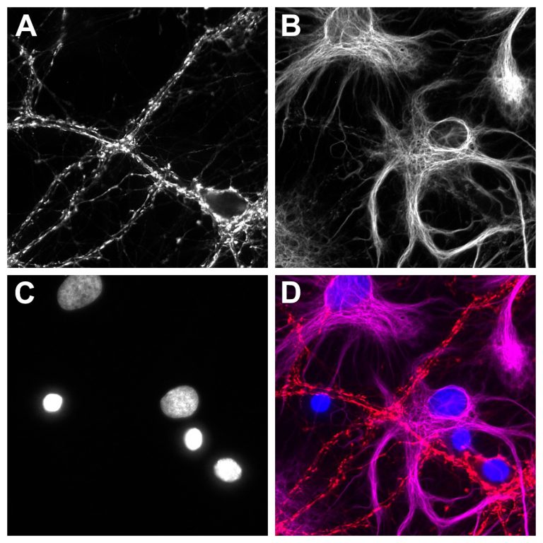
A fluorophore-conjugated single-domain antibody that specifically and strongly binds to the cytoplasmic domain of murine and rat Synaptotagmin 1 (SYT1).
FluoTag®-X2 anti-Synaptotagmin 1 (7D5) is derived from a single-domain antibody (sdAb) that specifically recognizes a synaptotagmin 1 (SYT1), a member of a protein family consisting of 17 members. Synaptotagmins are single-pass transmembrane proteins containing two calcium-binding domains in their intracellular portion. Synaptotagmin 1 is enriched at the presynaptic boutons on axon terminals, associated with synaptic vesicles, and is implicated in triggering vesicle fusion and neurotransmitter release upon a local increase in calcium concentration.
Our nanobody strongly and specifically binds to the cytosolic portion of mouse and rat SYT1.
FluoTag®-X2 anti-Synaptotagmin 1 (7D5) is directly conjugated to two fluorophores per sdAb (FluoTag®-X2 variant). To learn more about the FluoTags®, please visit our Technology section here.
| Variations: |
|
||||||||||||||||||||||||
|---|---|---|---|---|---|---|---|---|---|---|---|---|---|---|---|---|---|---|---|---|---|---|---|---|---|
| Related Products: | - | ||||||||||||||||||||||||
| Clone: | 7D5 | ||||||||||||||||||||||||
| Host: | Alpaca | ||||||||||||||||||||||||
| Produced in: | E.coli | ||||||||||||||||||||||||
| Application: |
IF Note: This Product is not recommended for detecting proteins in Western Blot, as sdAbs tend to recognize mainly native or folded proteins. |
||||||||||||||||||||||||
| Dilution: | 1:500 (corresponding to 5 nM final concentration) | ||||||||||||||||||||||||
| Capacity: | N/A | ||||||||||||||||||||||||
| Antigen: | - | ||||||||||||||||||||||||
| Targets: | Synaptotagmin 1 (cytosolic) | ||||||||||||||||||||||||
| Specificity: | Recognizes the cytosolic domain of rat and mouse Synaptotagmin 1 (other species not tested). | ||||||||||||||||||||||||
| Formulation: |
The single sdAb clone was lyophilized from PBS pH 7.4 containing 2% BSA (US-Origin). For more details, click the “Protocols” button above and check “Reconstitution and Storage”. |
||||||||||||||||||||||||
| kDa: | - | ||||||||||||||||||||||||
| Ext Coef: | - | ||||||||||||||||||||||||
| Shipping: | Ambient temperature | ||||||||||||||||||||||||
| Storing: |
Vials containing lyophilized reagent can be stored at 2-8°C for up to 12 months. After reconstitution, store at -80°C for up to 6 months. Working aliquots can be stored at -20°C for up to 4 weeks. For more details, click the “Protocols” button above and check “Reconstitution and Storage”. |
||||||||||||||||||||||||
| Protocols: |
Relevant protocols can be found under the “Protocols” button above. For additional information, visit our Resources page.
|
||||||||||||||||||||||||
| References: |
|
||||||||||||||||||||||||
| Notice: | To be used in vitro/ for research only. Non-toxic, non-hazardous, non-infectious. | ||||||||||||||||||||||||
| Legal terms: | By purchasing this product you agree to our general terms and conditions. |


