FluoTag®-X2 anti-GFAP
400,00 €
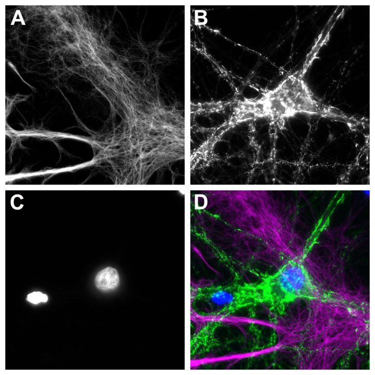
FluoTag-X2® anti-GFAP is a fluorescently conjugated single-domain antibody that recognizes mouse and rat GFAP. It carries two fluorophores and is equipped with specially designed linkers to work in expansion microscopy or enhance post-staining fixation for applications like dSTORM and DNA-PAINT.
FluoTag®-X2 anti-GFAP is derived from a single-domain antibody (sdAb) that specifically recognizes the Glial Fibrillary Acidic Protein (GFAP). GFAP belongs to type III intermediate filaments (like vimentin) and is expressed in various cell types throughout the human body. However, in the central nervous system, it is almost exclusively present in astrocytes, resulting in an important marker to differentiate neurons from astrocytes. GFAP is used primarily as an excellent astrocyte cellular marker.
Our nanobody binds selectively and strongly to mouse and rat GFAP.
FluoTag®-X2 anti-GFAP is directly conjugated to two fluorophores per sdAb (FluoTag®-X2 variant). Additionaly it is equipped with specially designed linkers to work in expansion microscopy or enhance post-staining fixation for applications like dSTORM and DNA-PAINT. To learn more about the FluoTags®, please visit our Technology section here.
| Variations: |
|
||||||||||||||||||||||||
|---|---|---|---|---|---|---|---|---|---|---|---|---|---|---|---|---|---|---|---|---|---|---|---|---|---|
| Related Products: | - | ||||||||||||||||||||||||
| Clone: | 1D12 | ||||||||||||||||||||||||
| Host: | Alpaca | ||||||||||||||||||||||||
| Produced in: | E.coli | ||||||||||||||||||||||||
| Application: | IF | ||||||||||||||||||||||||
| Dilution: | 1:500 (corresponding to 5 nM final concentration) | ||||||||||||||||||||||||
| Capacity: | N/A | ||||||||||||||||||||||||
| Antigen: | - | ||||||||||||||||||||||||
| Targets: | GFAP | ||||||||||||||||||||||||
| Specificity: | Recognizes murine and rat GFAP; other species or isoforms not yet tested. | ||||||||||||||||||||||||
| Formulation: | The single sdAb clone was lyophilized from PBS pH 7.4 containing 2% BSA (US-Origin). For more details, click the "Protocols" button above and check "Reconstitution and Storage". | ||||||||||||||||||||||||
| kDa: | - | ||||||||||||||||||||||||
| Ext Coef: | - | ||||||||||||||||||||||||
| Shipping: | Ambient temperature | ||||||||||||||||||||||||
| Storing: | Vials containing lyophilized reagent can be stored at 2-8°C for up to 12 months. After reconstitution, store at -80°C for up to 6 months. Working aliquots can be stored at -20°C for up to 4 weeks. For more details, click the "Protocols" button above and check "Reconstitution and Storage". | ||||||||||||||||||||||||
| Protocols: |
Relevant protocols can be found under the “Protocols” button above. For additional information, visit our Resources page.
|
||||||||||||||||||||||||
| References: |
|
||||||||||||||||||||||||
| Notice: | To be used in vitro/ for research only. Non-toxic, non-hazardous, non-infectious. | ||||||||||||||||||||||||
| Legal terms: | By purchasing this product you agree to our general terms and conditions. |


