
Immunostaining of PFA-fixed mouse hippocampus neurons with FluoTag®-X2 AZDye568 anti-PSD95 (Cat. No. N3702-AF568, dilution 1:500, represented in red) and FluoTag®-X2 Atto488 anti-GFAP (Cat. No. N3802-At488, dilution 1:500, represented in cyan). Nuclei were visualized by DAPI staining (gray). Figure modified from Kilisch et al., 2023 (see References below).

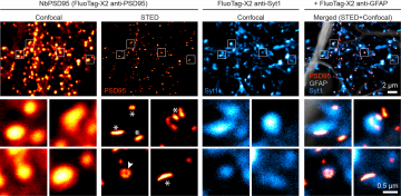
Confocal and STED images of primary hippocampal culture neurons stained using FluoTag®-X2 anti-PSD95 coupled to AberriorStar635P (Cat. No. N3702-Ab635P, dilution 1:500; displayed in hot red). Counterstaining performed with the FluoTag®-X2 anti-Syt1 (Cat. No. N2302-AF568, dilution 1:500; pre-synaptic marker displayed in hot cyan) and FluoTag®-X2 anti-GFAP (Cat. No. N3802-At488, dilution 1:500; glia marker displayed in gray). Four selected regions denoted by white squares are magnified, displaying conventional lateral PSD shapes (asterisks) and the perforated synapses (arrowheads) only recognizable under STED microscopy. Figure modified from Kilisch et al., 2023 (see References below).

IHC in mouse cerebellar cortex fixed in 4% formaldehyde for 24h and cut into 50 μm thick sections using a vibratome. Brightfield fluorescence imaging overview and the respective zoom-in (bottom) on the granular cell layer (G) and molecular layer (M). FluoTag®-X2 anti-PSD95 AbberiorStar635P (Cat. No. N3702-Ab635P, dilution 1:500; displayed in red) was used with recombinant anti-Synaptotagmin 1 antibody (Synaptic Systems) to reveal pre-synapses (cyan). DAPI staining in gray. FluoTag®-X2 anti-PSD95 gives a bright signal in the Purkinje cell layer at the axosomatic synapses, directly contacting the cell body of Purkinje cells (arrowheads). Figure modified from Kilisch et al., 2023 (see References below).


FluoTag®-X2 anti-PSD95
400,00 €
A fluorophore-conjugated single-domain antibody that specifically recognizes mouse and rat PSD95.
FluoTag®-X2 anti-PSD95 is derived from an in-house developed single-domain antibody (sdAb) that specifically recognizes a PSD95 (Postsynaptic Density Protein 95). PSD95 (alternative names: PSD-95, SAP-90 and DLG4) is a member of the MAGUK family, and it plays an essential role in forming a dynamic scaffold at the post-synapses. It functions as an organizing centrum of receptors, ion channels, kinases, and other relevant players in achieving synaptic plasticity and molecular changes during long-term potentiation.
Our nanobody binds selectively and strongly to one of the PDZ domains of PSD95, and it does not bind to either of its close family members, PSD93 or SAP102.
FluoTag®-X2 anti-PSD95 is directly conjugated to two fluorophores per sdAb (FluoTag®-X2 variant). To learn more about the FluoTags®, please visit our Technology section here.
| Variations: |
|
||||||||||||||||||||||||
|---|---|---|---|---|---|---|---|---|---|---|---|---|---|---|---|---|---|---|---|---|---|---|---|---|---|
| Related Products: | - | ||||||||||||||||||||||||
| Clone: | 1B2 | ||||||||||||||||||||||||
| Host: | Alpaca | ||||||||||||||||||||||||
| Produced in: | E.coli | ||||||||||||||||||||||||
| Application: | IF, IHC | ||||||||||||||||||||||||
| Dilution: | 1:500 (corresponding to 5 nM final concentration) , 5 µg/mL (IHC) | ||||||||||||||||||||||||
| Capacity: | N/A | ||||||||||||||||||||||||
| Antigen: | - | ||||||||||||||||||||||||
| Targets: | PSD95 | ||||||||||||||||||||||||
| Specificity: | Recognizes mouse and rat PSD95 (other species not tested). It does not cross-react with PSD93, SAP97 or SAP102. | ||||||||||||||||||||||||
| Formulation: | The single sdAb clone was lyophilized from PBS pH 7.4 containing 2% BSA (US-Origin). For more details, click the "Protocols" button above and check "Reconstitution and Storage". | ||||||||||||||||||||||||
| kDa: | - | ||||||||||||||||||||||||
| Ext Coef: | - | ||||||||||||||||||||||||
| Shipping: | Ambient temperature | ||||||||||||||||||||||||
| Storing: | Vials containing lyophilized reagent can be stored at 2-8°C for up to 12 months. After reconstitution, store at -80°C for up to 6 months. Working aliquots can be stored at -20°C for up to 4 weeks. For more details, click the "Protocols" button above and check "Reconstitution and Storage". | ||||||||||||||||||||||||
| Protocols: |
Relevant protocols can be found under the “Protocols” button above. For additional information, visit our Resources page. |
||||||||||||||||||||||||
| References: |
|
||||||||||||||||||||||||
| Notice: | To be used in vitro/ for research only. Non-toxic, non-hazardous, non-infectious. | ||||||||||||||||||||||||
| Legal terms: | By purchasing this product you agree to our general terms and conditions. |
Related products
-

FluoTag®-X2 anti-GFAP
Cat No: N3802FluoTag-X2® anti-GFAP is a fluorescently conjugated single-domain antibody that recognizes mouse and rat GFAP. It carries two fluorophores…
Select options -

Recombinant anti-PSD95 antibody (rHcAb)
The recombinant heavy-chain anti-PSD95 antibody is a genetic fusion of our established camelid anti-PSD95 single-domain antibody (sdAb) to…
Select options -

sdAb anti-PSD95
Cat No: N3705PSD95 (alternative names: PSD-95 or SAP-90) is a member of the MAGUK family, and it plays an important…
Select options