Recombinant anti-ALFA antibody (rHcAb)
420,00 €


The recombinant heavy-chain anti-ALFA antibody is a genetic fusion of our established camelid anti-ALFA single-domain antibody (sdAb) to various IgG Fc domains of the most commonly employed host species.
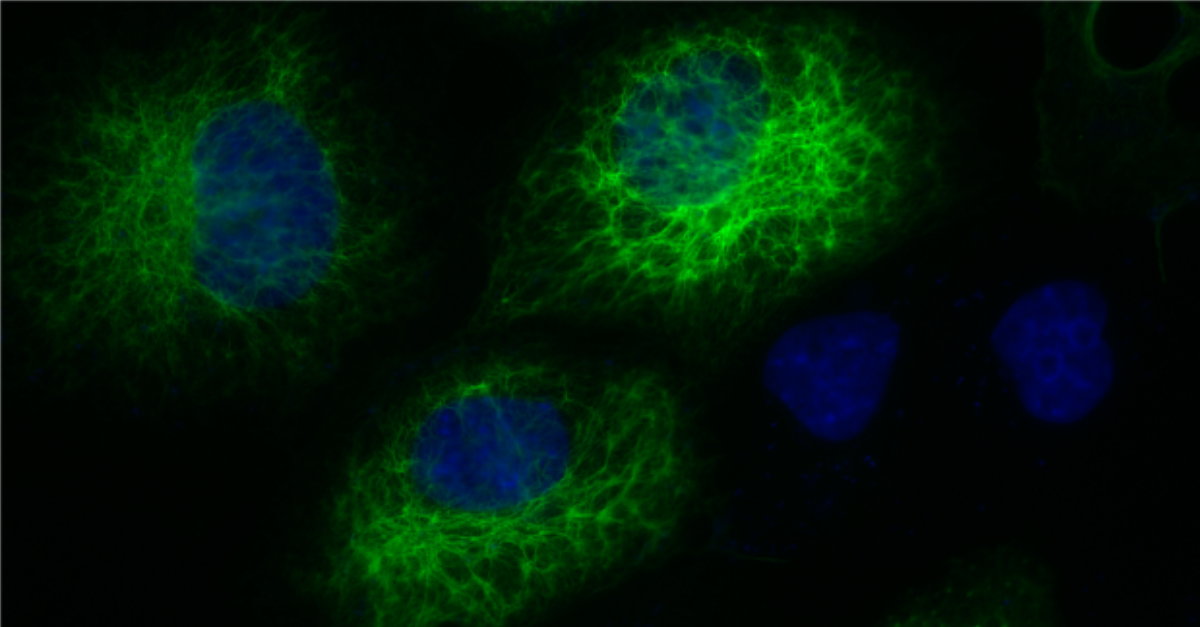
The recombinant heavy-chain anti-ALFA antibody is a genetic fusion of our established camelid anti-ALFA single-domain antibody (sdAb) to various IgG Fc domains of the most commonly employed host species. This antibody preserves the specificity and high-affinity binding of the anti-ALFA single-domain antibody and adds the features of a conventional IgG type antibody. It can be used in Western blotting and many other applications based on immunodetection (IF, IHC, IP). More information on recombinant heavy-chain antibodies (rHcAb) can be found here.
| Variations: |
|
||||||||||||||||||||||||
|---|---|---|---|---|---|---|---|---|---|---|---|---|---|---|---|---|---|---|---|---|---|---|---|---|---|
| Related Products: |
FluoTag®-X2 anti-ALFA (Cat. No. N1502) sdAb anti-ALFA (Cat. No. N1505) View related products |
||||||||||||||||||||||||
| Clone: | 1G5 | ||||||||||||||||||||||||
| Host: | Alpaca | ||||||||||||||||||||||||
| Produced in: | HEK | ||||||||||||||||||||||||
| Application: | IF, IHC, WB | ||||||||||||||||||||||||
| Dilution: | 1:500 (IF, IHC), 1:1000 WB | ||||||||||||||||||||||||
| Capacity: | N/A | ||||||||||||||||||||||||
| Antigen: | - | ||||||||||||||||||||||||
| Targets: | ALFA-tag | ||||||||||||||||||||||||
| Specificity: | Recognizes the ALFA-tag (SRLEEELRRRLTE). | ||||||||||||||||||||||||
| kDa: | - | ||||||||||||||||||||||||
| Ext Coef: | - | ||||||||||||||||||||||||
| Formulation: |
Lyophilized from PBS pH 7.4. Reconstitute with 100 µL 50% glycerol (v/v) in deionized water. If applicable, we recommend including 0.1% sodium azide as a preservative. For more details, click the Protocols button above and check Reconstitution and Storage. |
||||||||||||||||||||||||
| Shipping: | Ambient temperature | ||||||||||||||||||||||||
| Storage: |
Vials containing lyophilized reagent can be stored at 2-8°C for up to 12 months. After reconstitution, store at -80°C for up to 6 months. Working aliquots can be stored at -20°C for up to 4 weeks. For more details, click the Protocols button above and check Reconstitution and Storage. |
||||||||||||||||||||||||
| Protocols: |
Relevant protocols can be found under the Protocols button above. For additional information, visit our Resources page. |
||||||||||||||||||||||||
| References: |
|
||||||||||||||||||||||||
| Notice: | To be used in vitro/ for research only. Non-toxic, non-hazardous, non-infectious. | ||||||||||||||||||||||||
| Legal terms: |
By purchasing this product you agree to our general terms and conditions. |
||||||||||||||||||||||||
| Product LULL: | Limited Use Label License (LULL) – Internal Research Use Only applies. |
Related products
-

FluoTag®-X2 anti-ALFA
Cat No: N1502FluoTag®-X2 anti-ALFA is a fluorescently labeled single-domain antibody (sdAb) that specifically detects the small, rationally designed ALFA-tag. Enables…
Select options This product has multiple variants. The options may be chosen on the product page -

Affinity purified rabbit anti-ALFA polyclonal antibody
Cat No: N1581The anti-ALFA polyclonal antibody raised in rabbits recognizes the ALFA-tag (SRLEEELRRRLTE).
Add to cart -
Rabbit anti-ALFA polyclonal serum
Cat No: N1580The anti-ALFA polyclonal antibody raised in rabbit recognizes the ALFA-tag (SRLEEELRRRLTE).
Add to cart -

sdAb anti-ALFA
Cat No: N1505In-house developed single-domain antibody (sdAb) that recognizes the small rationally-designed ALFA-tag (SRLEEELRRRLTE) with low pM affinity.
Select options This product has multiple variants. The options may be chosen on the product page -

sdAb anti-ALFA Halo fusion
Cat No: N1541The sdAb anti-ALFA Halo fusion combines a single-domain antibody (sdAb) targeting the ALFA-tag with a dehalogenase-based technology. The…
Add to cart